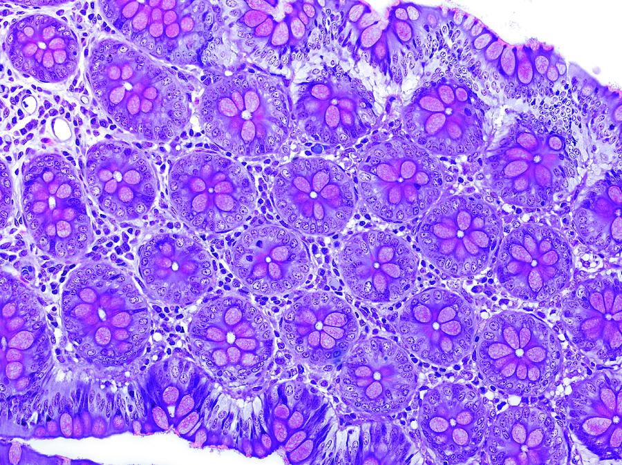
Small Intestine Tissue

by Microscape
Title
Small Intestine Tissue
Artist
Microscape
Medium
Photograph - Photograph
Description
Small intestine tissue. Light micrograph of a longitudinal section through tissue from the small intestine. This view shows cross-sections through many intestinal glands called crypts of Lieberkuhn. Crypts are long blind-ending tube-like extensions of the surface epithelial lining of the gut. In the small intestine they comprise several cell types including mucus-secreting goblet cells (purple) and absorptive enterocytes (blue) around a narrow central lumen. Crypts also contain gut epithelial stem cells. Connective tissue supporting the crypts contains fibroblasts, nerves, blood vessels, and white blood cells. Magnification: x183 when printed at 10 centimetres across.
Uploaded
July 20th, 2016
Embed
Share
Comments
There are no comments for Small Intestine Tissue. Click here to post the first comment.


