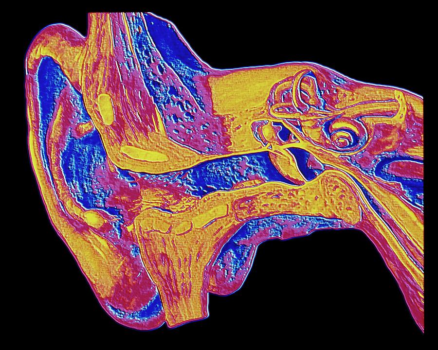
Computer Graphic Of The Anatomy Of The Human Ear

by Alfred Pasieka/science Photo Library
Title
Computer Graphic Of The Anatomy Of The Human Ear
Artist
Alfred Pasieka/science Photo Library
Medium
Photograph - Photograph
Description
Ear anatomy. Computer graphic of a section through the skull, showing the anatomy of the human ear. Sound waves are collected by the outer ear pinna (at left) and pass down the auditory canal to strike the eardrum (centre right). Vibrations of sound are transmitted via three tiny ear bones (malleus, incus and stapes) into fluid-filled structures of the inner ear (upper centre right). Three coils of the semi-circular canals detect balance and body orientation. While in the spiral-shaped cochlea, tiny hair cells are sensitive to vibrations of the cochlear fluid and these cells activate the auditory nerve (far right) which is connected to the brain.
Uploaded
September 24th, 2018
Statistics
Viewed 916 Times - Last Visitor from Fairfield, CT on 04/25/2024 at 12:00 PM
Embed
Share
Sales Sheet
Comments
There are no comments for Computer Graphic Of The Anatomy Of The Human Ear. Click here to post the first comment.


