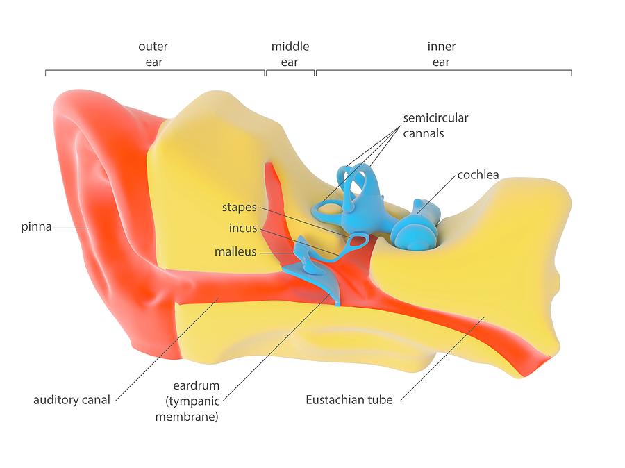
Human Ear Anatomy

by Science Photo Library
Title
Human Ear Anatomy
Artist
Science Photo Library
Medium
Photograph - Photograph
Description
Human ear anatomy. Illustration showing the outer (left), middle (centre) and inner (right) sections of a human ear. The outer ear's pinna (red, left) is the external part of the ear. The auditory canal (red) leads from here to the eardrum (tympanic membrane, blue), which transmits sounds from the air to the bones (blue) of the middle ear (malleus, incus, stapes). These mechanically transmit sounds to the fluid-filled organs (blue) of the inner ear, the semi-circular canals and the cochlea. Hair cells in the cochlea trigger nerve signals that are transmitted to the brain. The Eustachian tube (red, lower right) leads to the throat. For this artwork without labels, see C023/8842.
Uploaded
July 20th, 2016
Embed
Share
Comments
There are no comments for Human Ear Anatomy. Click here to post the first comment.


