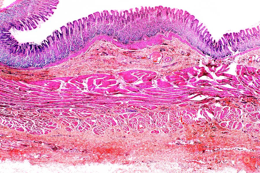
Stomach Wall

by Dr Keith Wheeler/science Photo Library
Title
Stomach Wall
Artist
Dr Keith Wheeler/science Photo Library
Medium
Photograph - Photograph
Description
Stomach wall. Light micrograph of a section through the stomach at the point of the fundus (top). The upper layer is the glandular mucosa, which has gastric pits (white areas between purple cells) where the gastric glands open to the digestive lumen (top). The light pink layer near top is the submucosa, which contains blood vessels (pink ovals, some with red blood cells in), lymph vessels and nerves. Beneath this are three layers of smooth muscle (bright pink), above the basement serous membrane (bottom). Magnification: x6 when printed at 10 centimetres wide.
Uploaded
October 8th, 2019
Embed
Share
Comments
There are no comments for Stomach Wall. Click here to post the first comment.


