Previous Image

|

Next Image
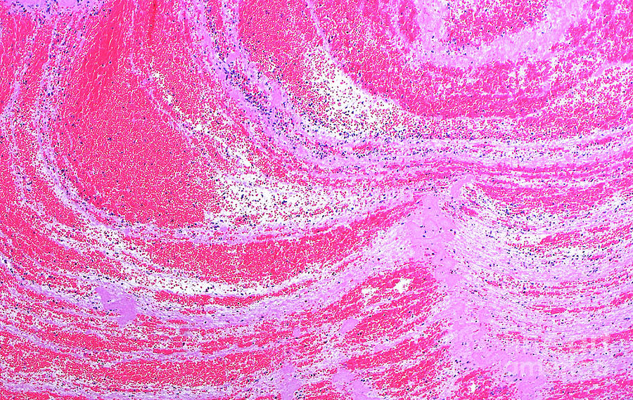
Blood Clot #1 Photograph by Ziad M. El-zaatari/science Photo Library

Similar Images

Nova View Automated Pathology Microscope by Aberration Films Ltd
Pathology by Tek Image
Pathology #1 by Tek Image
Pathology #2 by Tek Image
Pathology #3 by Tek Image
Pathology #4 by Tek Image
Pathology #5 by Tek Image
Pathology #6 by Tek Image
Nova View Automated Pathology Microscope #1 by Aberration Films Ltd
Forensic Pathology Laboratory by John Mclean/science Photo Library
Staining Pathology Slides by Choksawatdikorn / Science Photo Library
Staining Pathology Slides #1 by Choksawatdikorn / Science Photo Library
Staining Pathology Slides #2 by Choksawatdikorn / Science Photo Library
Staining Pathology Slides #3 by Choksawatdikorn / Science Photo Library
Staining Pathology Slides #4 by Choksawatdikorn / Science Photo Library
Preparing Bone Sections In Hospital Pathology Lab. by Simon Fraser/university Pathology Dept./ Rvi, Newcastle/science Photo Library
Preparing Bone Sections In Hospital Pathology Lab. #1 by Simon Fraser/university Pathology Dept./ Rvi, Newcastle/science Photo Library
Brain Slices Undergoing Gross Pathology by Simon Fraser/neuropathology Dept., Newcastle General Hospital/science Photo Library
Mrsa Ingestion By White Blood Cell #2 by Ami Images
Melanosis Coli by Cnri

Comments

There are no comments for Blood Clot #1.   Click here to post the first comment.

Recently Viewed

Blood Clot #1 by Ziad M. El-zaatari/science Photo Library
Previous Image Next Image

Help

Contact Us

Frequently Asked Questions

Return Policy

Returns

Terms of Use

Privacy Policy

Location Settings

Fairfield, CT (United States)

Currency: USD ($)

Units: Inches

Stay Connected

Sign up for our newsletter for exclusive deals, discount codes, and more.


Copyright © 2024 sciencephotogallery.com - All Rights Reserved - Website Powered by Fine Art America / Pixels - Original Source - Tapestries