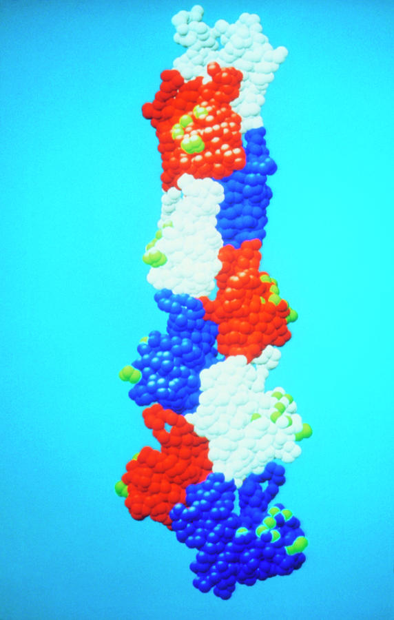
Model Of The Protein Actin

by Dr Kenneth Holmes/science Photo Library
Title
Model Of The Protein Actin
Artist
Dr Kenneth Holmes/science Photo Library
Medium
Photograph - Photograph
Description
Computer generated space-filling model of nine actin monomers from an F-actin helix, a polymer of the protein actin. For clarity each actin monomer (the repeated polymer unit) is shown in a different colour. Each amino-acid residue is represented by a sphere of radius 2.7 angstroms. In muscle cells, actin forms part of the thin filament, which cyclically interacts with the thick myosin filament to produce a mutual sliding that is the basis of muscle contraction. The green spheres are amino-acid residues that cross-link to the myosin in the actomyosin complex. The structure of the F-actin filament was determined using a technique called X-ray fibre diffraction.
Uploaded
September 15th, 2018
Statistics
Viewed 540 Times - Last Visitor from Syosset, NY on 04/26/2024 at 7:03 AM
Embed
Share
Sales Sheet
Comments
There are no comments for Model Of The Protein Actin. Click here to post the first comment.


