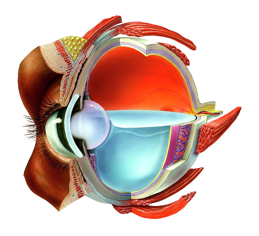
Eye Anatomy

by Bo Veisland/science Photo Library
Title
Eye Anatomy
Artist
Bo Veisland/science Photo Library
Medium
Photograph - Photograph
Description
Eye anatomy, artwork. At the front of the eye is the cornea (blue), a transparent coating. Behind this is the lens (opaque), which is partly covered by the iris (brown). The lens is held in place by zonular fibres attached to ciliary bodies (purple). The lens focuses light on the retina (yellow layer) at the back of the eye. Light sensitive cells in the retina transmit impulses to the brain via the optic nerve. Behind the retina is the choroid (purple), which contains blood vessels (blue) that nourish the back of the eye. The outer layer is the sclera, the white of the eye. The eyeball is filled with a gelatinous substance called vitreous humor. Several muscles hold the eyeball in place and allow it to rotate.
Uploaded
September 15th, 2018
Statistics
Viewed 888 Times - Last Visitor from Fairfield, CT on 04/24/2024 at 6:20 PM
Embed
Share
Sales Sheet
Comments
There are no comments for Eye Anatomy. Click here to post the first comment.


