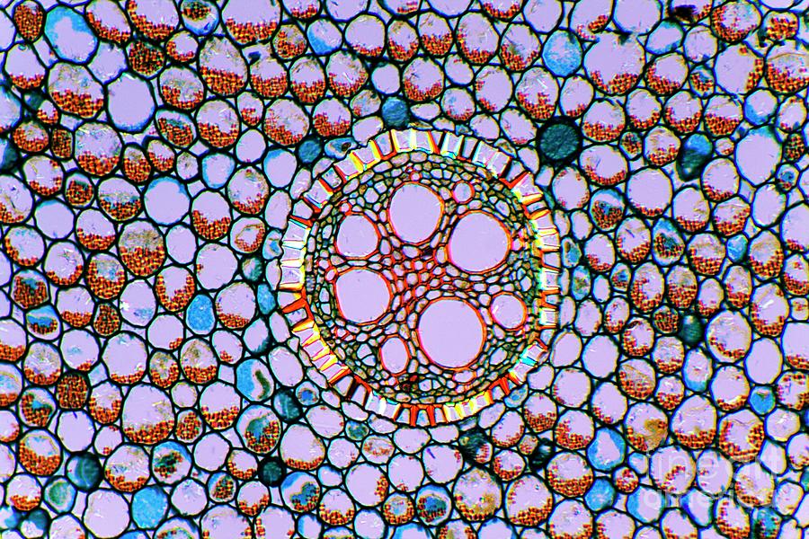
Iris Root #1

by Dr Keith Wheeler/science Photo Library
Title
Iris Root #1
Artist
Dr Keith Wheeler/science Photo Library
Medium
Photograph - Photograph
Description
Iris root. Polarised light micrograph of a section through the root of an Iris plant (Iris germanica), showing a vascular cylinder (centre) and parenchyma cells packed with starch grains. The cylinder is comprised of a central cluster of parenchyma cells (red) surrounded by vascular bundles (brown) and the endodermis (multicoloured ring). The largest vessels seen here are metaxylem, part of the xylem tissue (orange) in the vascular bundles. Xylem transports water and mineral nutrients from the roots throughout the plant, while the phloem (brown/dark green), the other component of the bundles, transports carbohydrates and plant hormones. Magnification: x100 when printed 10 centimetres wide.
Uploaded
November 13th, 2019
Statistics
Viewed 577 Times - Last Visitor from New York, NY on 04/20/2024 at 3:55 AM
Embed
Share
Sales Sheet
Comments
There are no comments for Iris Root #1. Click here to post the first comment.


