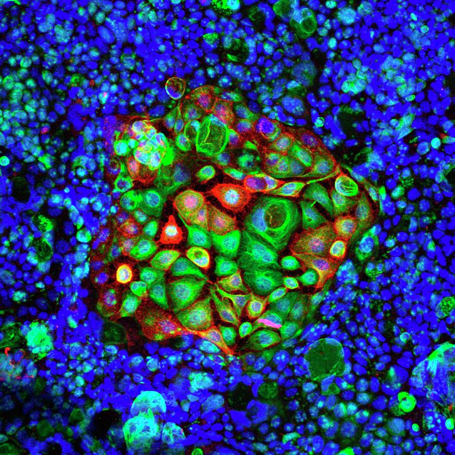
Colorectal Cancer Cells

by Ammrf, University Of Sydney
Title
Colorectal Cancer Cells
Artist
Ammrf, University Of Sydney
Medium
Photograph - Photograph
Description
Colorectal cancer cells. Confocal light micrograph of cultured colorectal cancer cells dividing. The cellular proteins are indicated by fluorescent markers: DAPI (blue, cell nuclei), tubulin (green), GM1 (red). The central island of flat cells (green) contains dividing cells and is surrounded by differentiated cells. The cells in the island have unusual properties and can potentially be used to study the effectiveness of new anti-cancer drugs. Magnification: x160 when printed at 10 centimetres across.
Uploaded
July 6th, 2016
Statistics
Viewed 1,116 Times - Last Visitor from Ottawa, ON - Canada on 04/25/2024 at 12:31 AM
Embed
Share
Sales Sheet


