
Boundary: Bleed area may not be visible.


by Dr Lothar Schermelleh
$41.95
Model
Case Style
Orientation
Image Size
Product Details
Cell Division iPhone case by Dr Lothar Schermelleh. Protect your iPhone with an impact-resistant, slim-profile, hard-shell case. The image is printed directly onto the case and wrapped around the edges for a beautiful presentation. Simply snap the case onto your iPhone for instant protection and direct access to all of the phone's features!
Design Details
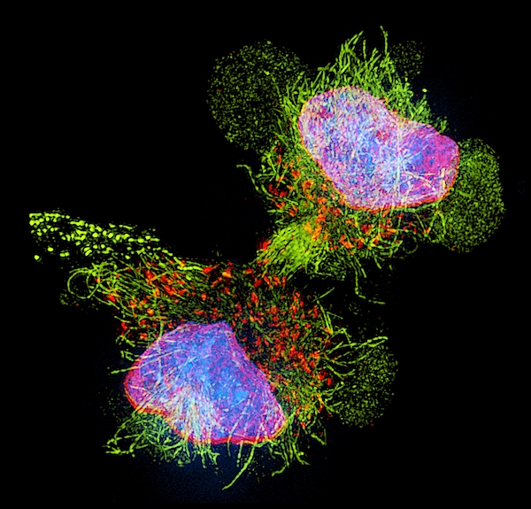
Cell division. 3D-structured illumination micrograph of the nucleus of a mouse myoblast cell during cytokinesis (cell division). Cytokinesis occurs... more
Ships Within
3 - 4 business days
Protect your with an impact-resistant, slim-profile, hard-shell case. The image is printed directly onto the case and wrapped around the edges for a beautiful presentation. Simply snap the case onto your for instant protection and direct access to all of the phone's features!
Cell division. 3D-structured illumination micrograph of the nucleus of a mouse myoblast cell during cytokinesis (cell division). Cytokinesis occurs after nuclear division (mitosis), which produces two daughter nuclei (blue and pink) from one parent nuclei. Here the nuclear envelopes (red) are reforming around the chromatin (blue), the complex of DNA (deoxyribonucleic acid) and protein that forms chromosomes. Microtubules, which moved the DNA to the new nuclei, are yellow.
Science Photo Library (SPL) is the leading source of science images and footage. Sourced from scientific and medical experts, acclaimed photographers and renowned institutions, our content is unrivaled worldwide. Outstanding quality, accuracy and commitment to excellence are deeply embedded in our DNA. Science Photo Library inspires creative professionals and delivers engaging content of the highest quality for a wide range of clients in a variety of sectors. Visit sciencephoto.com for more information and stay connected on Twitter, LinkedIn, Instagram and Vimeo.
$41.95


Copyright © 2024 sciencephotogallery.com - All Rights Reserved - Website Powered by Fine Art America / Pixels - Original Source - Tapestries


Sunil Kapadia
Congratulations !