Previous Image

|

Next Image
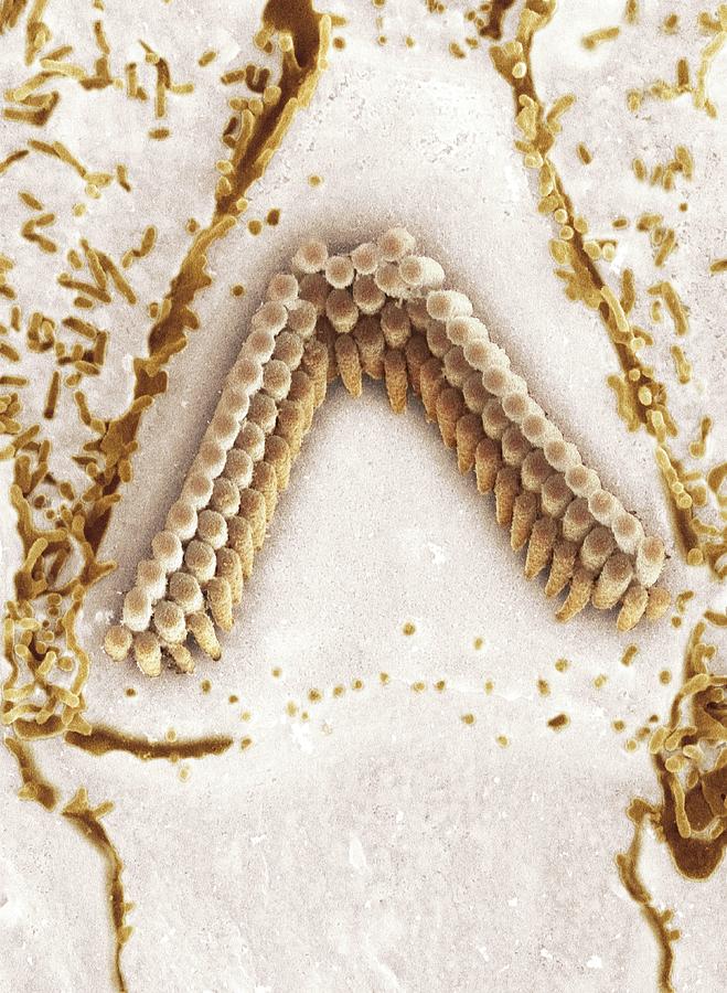
Inner Ear Hair Cells, Sem #1 Photograph by Dr David Furness, Keele University

Similar Images

Sensory Hair Cells In Ear #1 by Susumu Nishinaga
Damaged Inner Ear Hair Cells by Dr Goran Bredberg/science Photo Library
Normal Inner Ear Hair Cells by Dr Goran Bredberg/science Photo Library
Illustration Of Human Cilia by Sebastian Kaulitzki/science Photo Library
Illustration Of Human Cilia #1 by Sebastian Kaulitzki/science Photo Library
Illustration Of Human Cilia #2 by Sebastian Kaulitzki/science Photo Library
Illustration Of Human Cilia #3 by Sebastian Kaulitzki/science Photo Library
Illustration Of Human Cilia #4 by Sebastian Kaulitzki/science Photo Library
Illustration Of Human Cilia #5 by Sebastian Kaulitzki/science Photo Library
Cilia by Jose Calvo / Science Photo Library
Cilia And Microvilli by Jose Calvo / Science Photo Library
Cilia #1 by Jose Calvo / Science Photo Library
Axoneme Microtubule Structure by Ramon Andrade 3dciencia/science Photo Library
Paramecium Ciliates #1 by Gerd Guenther/science Photo Library
Squantinella Rotifer by Gerd Guenther/science Photo Library
Coloured Sem Of Fallopian Tube Cilia & Microvilli by Dr Yorgos Nikas/science Photo Library
Stomach Cilia #2 by Marian Miller
Fallopian Tube #3 by Steve Gschmeissner
Ciliate Protozoan by Steve Gschmeissner
Vorticella by Steve Gschmeissner

Comments

There are no comments for Inner Ear Hair Cells, Sem #1.   Click here to post the first comment.

Recently Viewed

Inner Ear Hair Cells, Sem #1 by Dr David Furness, Keele University
Previous Image Next Image

Help

Contact Us

Frequently Asked Questions

Return Policy

Returns

Terms of Use

Privacy Policy

Location Settings

Cambridge, MA (United States)

Currency: USD ($)

Units: Inches

Stay Connected

Sign up for our newsletter for exclusive deals, discount codes, and more.


Copyright © 2024 sciencephotogallery.com - All Rights Reserved - Website Powered by Fine Art America / Pixels - Original Source - Tapestries