
Boundary: Bleed area may not be visible.

Inside Message (Optional)
Inside View

by Jose Calvo / Science Photo Library
$5.00
Quantity
The more you buy... the more you save.
Orientation
Image Size
Product Details
Our greeting cards are 5" x 7" in size and are produced on digital offset printers using 100 lb. paper stock. Each card is coated with a UV protectant on the outside surface which produces a semi-gloss finish. The inside of each card has a matte white finish and can be customized with your own message up to 500 characters in length. Each card comes with a white envelope for mailing or gift giving.
Design Details
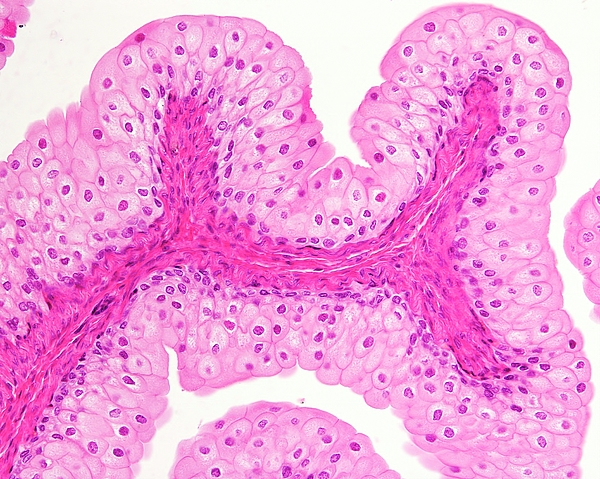
Light micrograph of one of the mucosal folds that appear in an empty urinary bladder. The axis of the fold is formed by connective tissue of the... more
Ships Within
2 - 3 business days
Light micrograph of one of the mucosal folds that appear in an empty urinary bladder. The axis of the fold is formed by connective tissue of the lamina propria. This axis is covered by a transitional epithelium, also called urothelium, highly specific to the urinary tract. In the empty bladder, the epithelium is multi-layered showing 4 or 5 layers. The surface cells are large, bulging into the bladder lumen. Magnification: x180 when printed at 10 centimetres across.
Science Photo Library (SPL) is the leading source of science images and footage. Sourced from scientific and medical experts, acclaimed photographers and renowned institutions, our content is unrivaled worldwide. Outstanding quality, accuracy and commitment to excellence are deeply embedded in our DNA. Science Photo Library inspires creative professionals and delivers engaging content of the highest quality for a wide range of clients in a variety of sectors. Visit sciencephoto.com for more information and stay connected on Twitter, LinkedIn, Instagram and Vimeo.
$5.00


Copyright © 2024 sciencephotogallery.com - All Rights Reserved - Website Powered by Fine Art America / Pixels - Original Source - Tapestries


There are no comments for Urinary Bladder Epithelium. Click here to post the first comment.