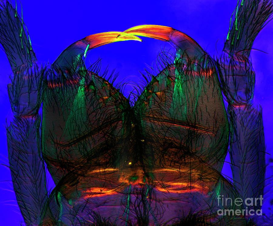
Spider Mouthparts

by Dr Keith Wheeler/science Photo Library
Title
Spider Mouthparts
Artist
Dr Keith Wheeler/science Photo Library
Medium
Photograph - Photograph
Description
Spider mouthparts. Polarised light micrograph of the chelicerae and pedipalps of a water spider (Argyroneta aquatica). The jointed pedipalps are on both sides of the central two chelicerae, which have jointed fang-like tips (orange-yellow). A duct from the poison gland opens just below the inner ends of the fangs. The chelicerae stab inwards to pierce the prey and then inject poison which kills them. The mouth, below the bases of the chelicerae, then injects saliva with digestive enzymes, which breaks down the inside of the victim. The resulting juices are then sucked up. Magnification: x36 when printed at 10 centimetres across.
Uploaded
October 16th, 2019
Statistics
Viewed 440 Times - Last Visitor from Ottawa, ON - Canada on 04/25/2024 at 1:59 AM
Embed
Share
Sales Sheet
Comments
There are no comments for Spider Mouthparts. Click here to post the first comment.


