Intestinal Villi

by Astrid & Hanns-frieder Michler/science Photo Library
Title
Intestinal Villi
Artist
Astrid & Hanns-frieder Michler/science Photo Library
Medium
Photograph - Photograph
Description
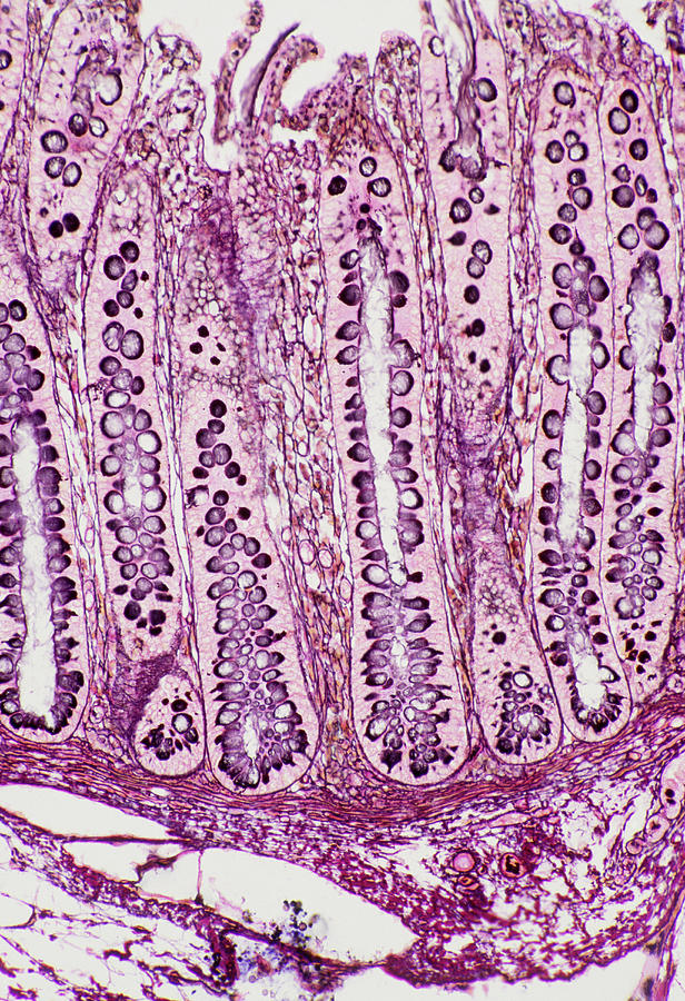
Intestinal villi. Light micrograph of a section through villi on the lining of the large intestine. The folds of the wall of the large intestine are covered with these small projections called villi, which serve to increase the surface area for the absorption of water from digested food. Villi are mainly composed of columnar epithelial cells, whose nuclei (dark purple) are revealed by the stain used. The bases of the villi are across bottom, and the lumen (the internal surface of the intestine) is across top. Comori- Azan stain. Magnification: x140 when printed 10cm high.
Uploaded
September 28th, 2018
Statistics
Viewed 384 Times - Last Visitor from New York, NY on 04/21/2024 at 12:36 PM
Embed
Share
Sales Sheet
Comments
There are no comments for Intestinal Villi. Click here to post the first comment.


