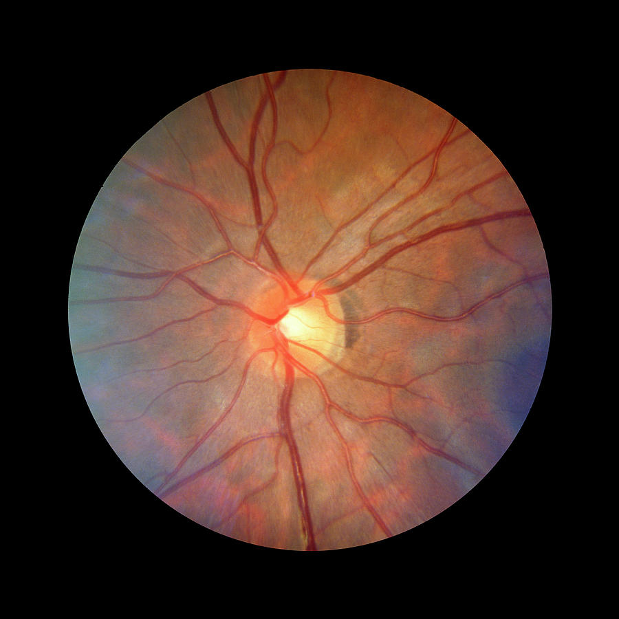
Fundus Camera Image Of A Normal Retina

by Rory Mcclenaghan/science Photo Library
Title
Fundus Camera Image Of A Normal Retina
Artist
Rory Mcclenaghan/science Photo Library
Medium
Photograph - Photograph
Description
Fundus camera image of the retina of a normal eye, showing the distribution of the retinal veins & arteries. The central retinal artery enters the optic nerve before it reaches the eyeball, and emerges from the centre of the optic disc (the blind spot of the eye, the pale central area). On the right can be seen the dark edge of the macula, a region free of large blood vessels where the fovea (the area of highest visual acuity) is found. An extension of the optic nerve, the retina consists of photosensitive cells (rods & cones) which translate light energy into nervous impulses. This retina has a greenish hue because the subject was Asian. In Caucasians it is reddish
Uploaded
September 13th, 2018
Statistics
Viewed 1,107 Times - Last Visitor from New York, NY on 04/15/2024 at 12:10 PM
Embed
Share
Sales Sheet
Comments
There are no comments for Fundus Camera Image Of A Normal Retina. Click here to post the first comment.


