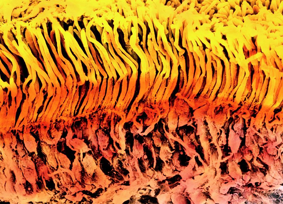
Coloured Sem Of A Section Through The Human Retina

by Photo Insolite Realite & V. Gremet/science Photo Library
Title
Coloured Sem Of A Section Through The Human Retina
Artist
Photo Insolite Realite & V. Gremet/science Photo Library
Medium
Photograph - Photograph
Description
Retinal cells. Coloured Scanning Electron Micrograph (SEM) of a section through the human retina. The retina is the light-sensitive layer at the back of the eye. Light sensitive cells are known as rods and cones, and these cells are long and yellow (at upper image). At lower image (pink) are the bodies of nerve cells which transmit impulses from the rods and cones to the brain via the optic nerve. The surface of the retina, from where light enters this layer, is at bottom of image (not seen). Rods contain a pigment, rhodopsin, which is broken down by light and transmits a signal to nerve cells. Cone cells detect colour. Magnification: x1400 when printed 10cm wide.
Uploaded
September 23rd, 2018
Statistics
Viewed 1,003 Times - Last Visitor from Bradford, West Yorkshire - United Kingdom on 04/23/2024 at 7:11 AM
Embed
Share
Sales Sheet


