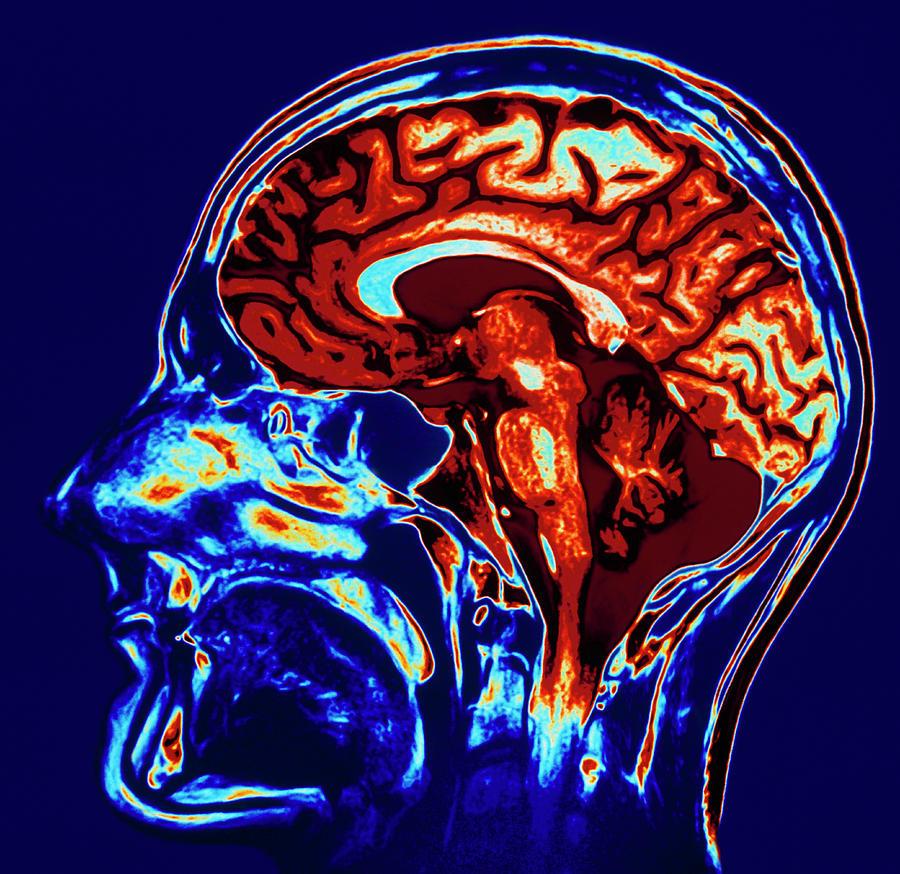
Coloured Mri Scan Of Brain In Sagittal Se

by Geoff Tompkinson
Title
Coloured Mri Scan Of Brain In Sagittal Se
Artist
Geoff Tompkinson
Medium
Photograph - Photograph
Description
Healthy brain. Coloured Magnetic Resonance Imaging (MRI) scan of a healthy human brain seen in a sagittal section. The large cerebrum is seen as a folded orange mass at upper frame. This is where all the brain's 'higher thought activities' occur. The two hemispheres that make up the cerebrum are connected by a thick band of nerve fibres known as the corpus callosum, seen as a blue band at upper centre. At centre right is the brown-triangular cerebellum which coordinates muscle action, balance and posture. The brainstem, a stalk of nerve tissue connecting the brain with the spinal cord, is seen at centre.
Uploaded
October 7th, 2018
Statistics
Viewed 767 Times - Last Visitor from Cambridge, MA on 04/16/2024 at 4:57 PM
Embed
Share
Sales Sheet
Comments
There are no comments for Coloured Mri Scan Of Brain In Sagittal Se. Click here to post the first comment.


