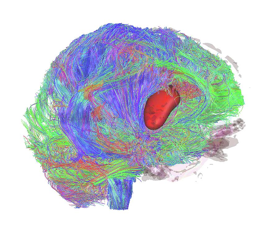
Brain Tumour

by Sherbrooke Connectivity Imaging Lab/science Photo Library
Title
Brain Tumour
Artist
Sherbrooke Connectivity Imaging Lab/science Photo Library
Medium
Photograph - Photograph
Description
Brain tumour. 3D diffusion tensor imaging (DTI) magnetic resonance imaging (MRI) scan of nerve pathways (coloured) in a brain with a tumour (red, centre right). The brain is seen from the side, with the front of the brain at right. Brain tumours can be benign or malignant (cancerous). They can cause seizures, headaches, and memory and personality changes due to the growth of the tumour. The nerve fibres include the corticospinal tract (blue), passing from the motor cortex to the spinal cord. Diffusion tensor imaging (tractography) measures the direction of water diffusion, which in the brain reveals the orientation of nerve fibres.
Uploaded
October 7th, 2018
Statistics
Viewed 701 Times - Last Visitor from Fairfield, CT on 04/24/2024 at 12:36 AM
Embed
Share
Sales Sheet


