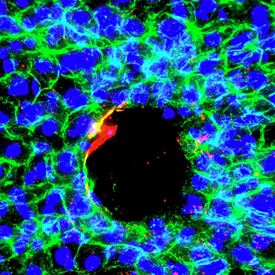
Liver Tissue #6

by R. Bick, B. Poindexter, Ut Medical School/science Photo Library
Title
Liver Tissue #6
Artist
R. Bick, B. Poindexter, Ut Medical School/science Photo Library
Medium
Photograph - Photograph
Description
Liver tissue. Fluorescence deconvolution micrograph of a section through liver tissue. This tissue sample includes a cross-section through one of the liver's central veins (black). These veins are the central part of a liver lobule, receive the blood that has circulated through the liver, returning it to the body. The liver stores nutrients and helps to clean the blood of toxins and other waste products. Cellular proteins are highlighted with fluorescent markers: smooth muscle actin (red), f-actin (green) and cell nuclei (blue).
Uploaded
October 7th, 2018
Statistics
Viewed 514 Times - Last Visitor from Fairfield, CT on 04/19/2024 at 7:10 PM
Embed
Share
Sales Sheet
Comments
There are no comments for Liver Tissue #6. Click here to post the first comment.


