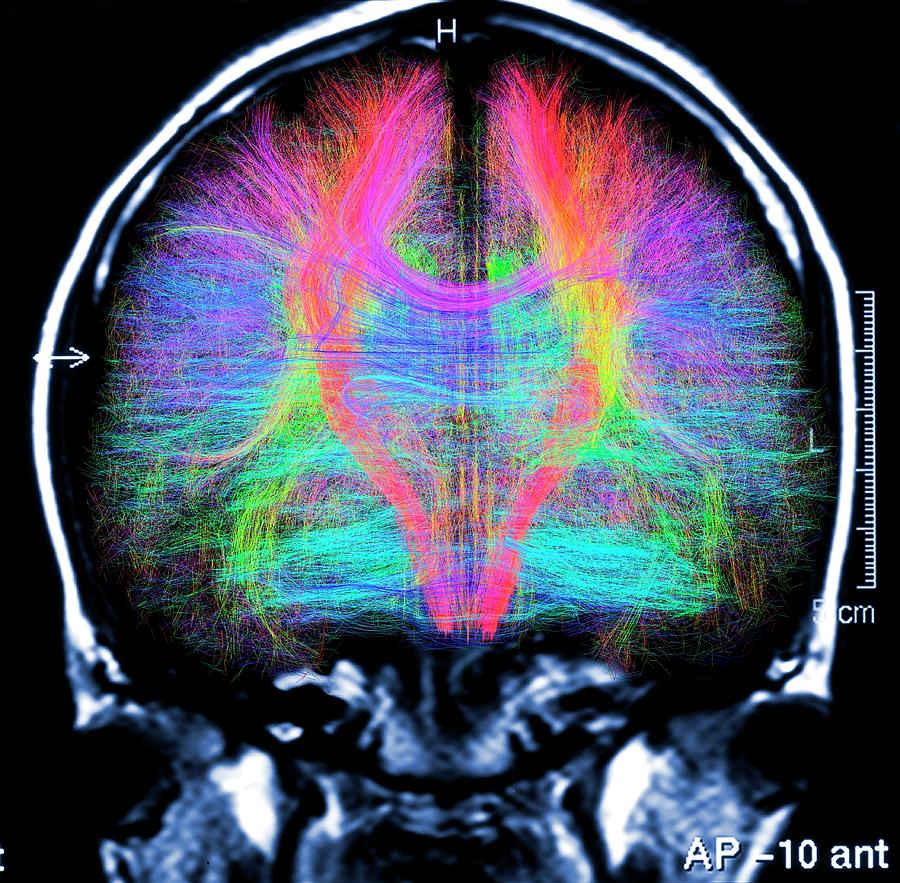
Brain Mri And White Matter Fibres #3

by Alfred Pasieka/science Photo Library
Title
Brain Mri And White Matter Fibres #3
Artist
Alfred Pasieka/science Photo Library
Medium
Photograph - Photograph
Description
Computer artwork of a MRI frontal view of the brain showing white matter fibres. Coloured 3D diffusion spectral imaging (DSI) scan of the bundles of white matter nerve fibres in the brain. The fibres transmit nerve signals between brain regions and between the brain and the spinal cord. Diffusion spectrum imaging (DSI) is a variant of magnetic resonance imaging (MRI) in which a magnetic field maps the water contained in neuron fibers, thus mapping their criss-crossing patterns. A similar technique called diffusion tensor imaging (DTI) is also used to explore neural data of white matter fibres in the brain. Both methods allow mapping of their orientations and the connections between brain regions. Data/software: NIH Human Connectome Project / www.humanconnectomeproject.org)
Uploaded
September 15th, 2018
Statistics
Viewed 1,755 Times - Last Visitor from New York, NY on 04/24/2024 at 4:33 AM
Embed
Share
Sales Sheet


